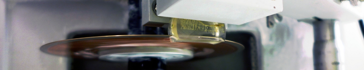
MMLAB steht für „Interfacultary Laboratory for Micro- and Nanomechanics of Biological and Biomimetical Materials“. Das Labor befindet sich an der Technischen Universität Wien und konzentriert sich auf die Untersuchung der mechanischen und morphologischen Eigenschaften von Mikro- und Nanostrukturen. Besonderer Wert wird auf biologisches Material wie verkalktes Gewebe und Holz gelegt. Zu den experimentellen Mitteln gehören:
- Nanoindentierung
- Rastersondenmikroskopie
- Mikro Computertomographie
- (polarisierte und fluoreszierende) Lichtmikroskopie
- Zug-/Torsionsprüfung
- Optische Bewegungserfassung
- Hochgeschwindigkeitsvideo
Die Probenvorbereitung wird in einem Vorbereitungsraum durchgeführt, der speziell für biologisches Gewebe ausgestattet ist.
Das MMLAB ist interfakulär und gehört zu den drei verschiedenen Instituten (in alphabetischer Reihenfolge):
- Institut für Leichtbau und Struktur-Biomechanik
- Institut für Werkstoffwissenschaft und Werkstofftechnologie
- Institut für Mechanik der Werkstoffe und Strukturen
Das Labor wird verwendet für Auftragsforschung, Industrieprojekte und Wissenschaftliche Kooperationen.